Much of the discussion around New Approach Methods (NAMs) and animal models has been about total replacement, but many researchers in both fields recognize that we are not yet at a place where animal models in biomedical research can be eliminated entirely. Cancer research is one of many fields that rely on both models, and researchers in Oregon are using them side by side.
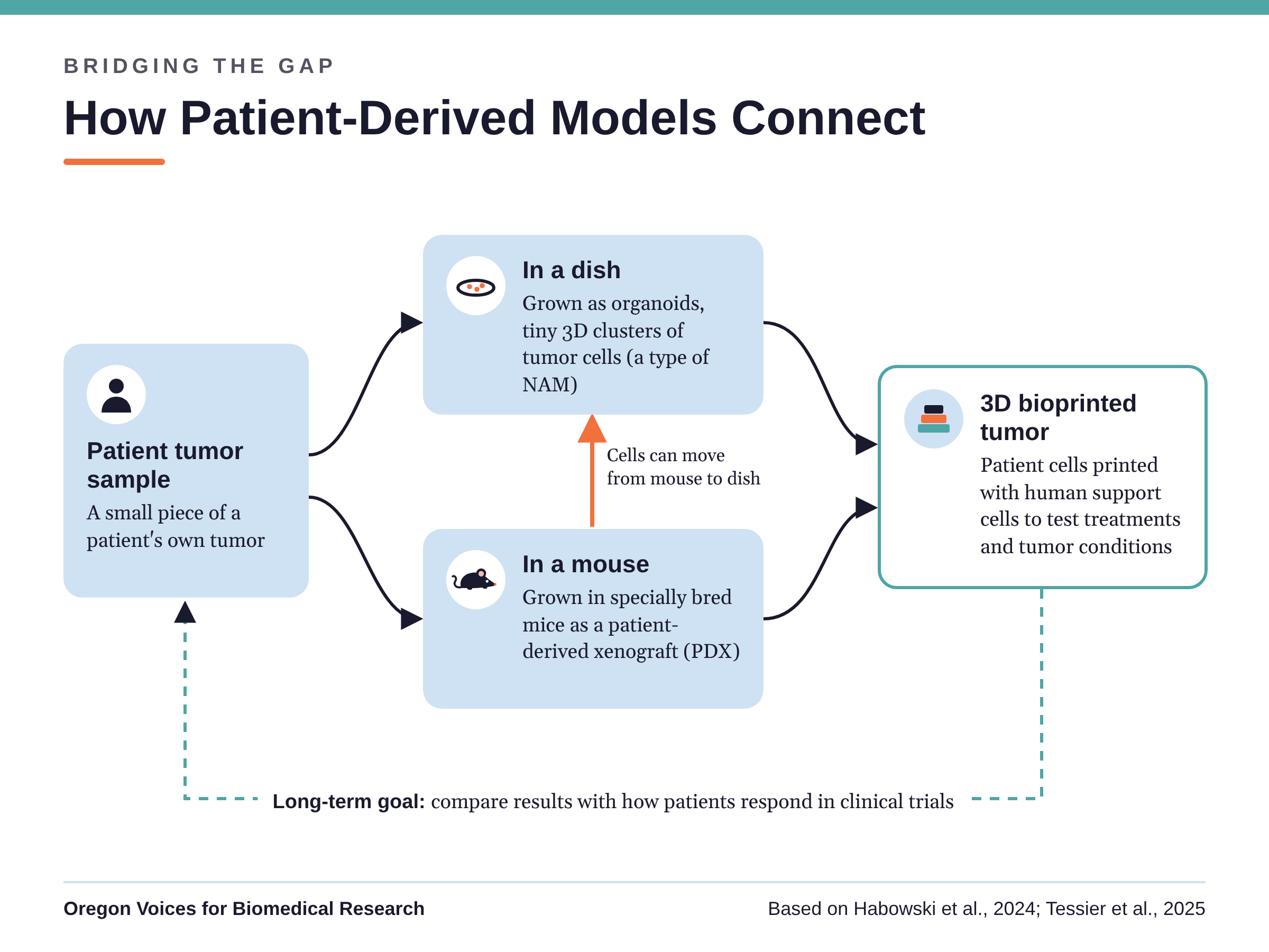
One way they do this is with patient-derived cancer models. Scientists start with a small sample of a patient’s own tumor, which can be grown in the lab as tiny 3D clusters of cells called “organoids” (a type of NAM), or placed into specially bred mice with immune systems that won’t reject the human tumor, creating a patient-derived xenograft (PDX). Each model can answer questions the other can’t, and by using them together researchers can get a fuller picture of how a tumor grows and responds to treatment.
Three scientists at Oregon Health & Science University’s (OHSU) Knight Cancer Institute are part of a national patient-derived cancer model consortium supported by the National Cancer Institute (NCI). In 2024, they co-authored a paper sharing what the consortium has learned about choosing and using these models. The consortium paper is built around 10 short “vignettes,” each written by one of the member research groups. In this post, we’ll look at the Oregon researchers behind the paper and how their work connects to it, along with the consortium’s advice for choosing the right model.
Meet Oregon’s Consortium Members
Drs. Rosalie Sears, Ellen Langer, and Jonathan Brody are all members of the OHSU Knight Cancer Institute and the Brenden-Colson Center for Pancreatic Care, and pancreatic cancer runs through much of their work. Pancreatic ductal adenocarcinoma is one of the most lethal cancers, and standard treatments have done little to improve how well patients do (Habowski et al., 2024). All three also collaborated on a 2023 study where they packaged a potential cancer drug called juglone, which has been held back by concerns about its toxicity, into tiny carrier particles, testing it first on established cancer cell lines in a dish and then in mice, where the new formulation slowed tumor growth without the toxicity problems (Shah et al., 2023).
Rosalie Sears
Dr. Sears led a 2019 study, with Langer as first author, that used 3D bioprinting to build small pieces of breast and pancreatic tumor tissue in the lab, including some made from patients’ own tumor tissue. The team showed that these printed tissues could recreate aspects of real tumors, giving researchers a way to study how cancer cells interact with the cells around them (Langer et al., 2019). Sears also co-authored a step-by-step method in 2025, from a team at OHSU, for taking human pancreatic cancer cells out of patient-derived xenografts and growing them in culture (Tessier et al., 2025). This is the step that connects the mouse and the dish, allowing the two models to work together.
Ellen Langer
Along with her role in the 2019 bioprinting study, Dr. Langer has continued working to improve organoid models. In a 2025 review, her team looked at how the gels and nutrient mixtures used to grow gastrointestinal organoids can change how the cells behave and respond to treatment, and how better growing conditions could make organoids more useful (Kuhn et al., 2025). The review presents organoids as an encouraging way to study how human cells work, one that could serve in place of both flat cell cultures and animal models. Langer has also co-authored mouse studies, which is a good example of the same scientists advancing both kinds of models.
Jonathan Brody
Dr. Brody, who is also part of OHSU’s Department of Surgery, focuses on finding better treatments for pancreatic cancer. In a 2024 study, his team tested CF10, a newer relative of a standard pancreatic cancer chemotherapy drug, called 5-FU, that can cause serious side effects. In lab tests, CF10 was more than 300 times as potent as 5-FU and worked at very low doses in models grown from patients’ own tumors (Finan et al., 2024). The team noted that the drug combinations they identified still need to be tested in living animals, which follows the consortium’s recommended approach of testing in vitro (in cell cultures) before moving to animals.
Where Their Work Meets the Consortium Paper
One of the consortium’s vignettes discusses the use of 3D bioprinting to recreate pancreatic tumor tissue, and it cites the 2019 OHSU bioprinting study along with four pancreatic cancer studies Brody co-authored (Habowski et al., 2024). The paper doesn’t say which member group wrote each vignette, so we can’t point to it as Oregon’s alone, but the overlap with the OHSU team’s published work is clear.
The vignette describes the same path from patient to mouse to dish covered earlier. A patient’s cancer cells are first grown in larger numbers, either in a dish or in mice as a patient-derived xenograft, and then printed into small tissues surrounded by human support cells and immune cells. Each tissue takes about a minute to print and can be kept alive for more than three weeks. Because researchers control every ingredient, they can test how conditions around a tumor, like glucose, oxygen, or magnesium levels, affect how well a drug works (Habowski et al., 2024). Studying these kinds of interactions one factor at a time is difficult in living tissue, which is part of what makes printed tumors useful (Langer et al., 2019). The authors’ long-term goal is to compare results from these printed tumors with how patients in clinical trials actually respond, which is how they’ll learn how much these models can truly predict outcomes.
Choosing the Right Tool for the Question
Beyond the individual vignettes, the consortium authors offer advice that applies to any lab working with these models. Their main recommendation is to start with the simplest and least expensive model that can answer the research question, and to move to more complex systems, such as going from organoids to mice, only when the simpler model can’t provide the answer (Habowski et al., 2024). They also point out that having a model available isn’t, by itself, a reason to use it, since the strongest science comes from matching the model to the question.
This lines up closely with the 3Rs, particularly Reduction, which aims to use the fewest animals needed to get reliable results. Animal models generally cost more and take longer than in vitro models (Habowski et al., 2024), so researchers have practical reasons, in addition to ethical ones, to save them for the questions that truly need them.
A known drawback of patient-derived xenografts is that, because the mice are bred with immune systems that won’t reject a human tumor (Habowski et al., 2024), they can’t fully show how a person’s immune system would fight the cancer, an important issue for immunotherapy research. To address this, some researchers use “humanized” mice, which receive human blood-forming stem cells so they develop parts of a human immune system. The consortium authors acknowledge that humanized mice are expensive and slow to develop, but say certain questions can’t be answered without them (Habowski et al., 2024).
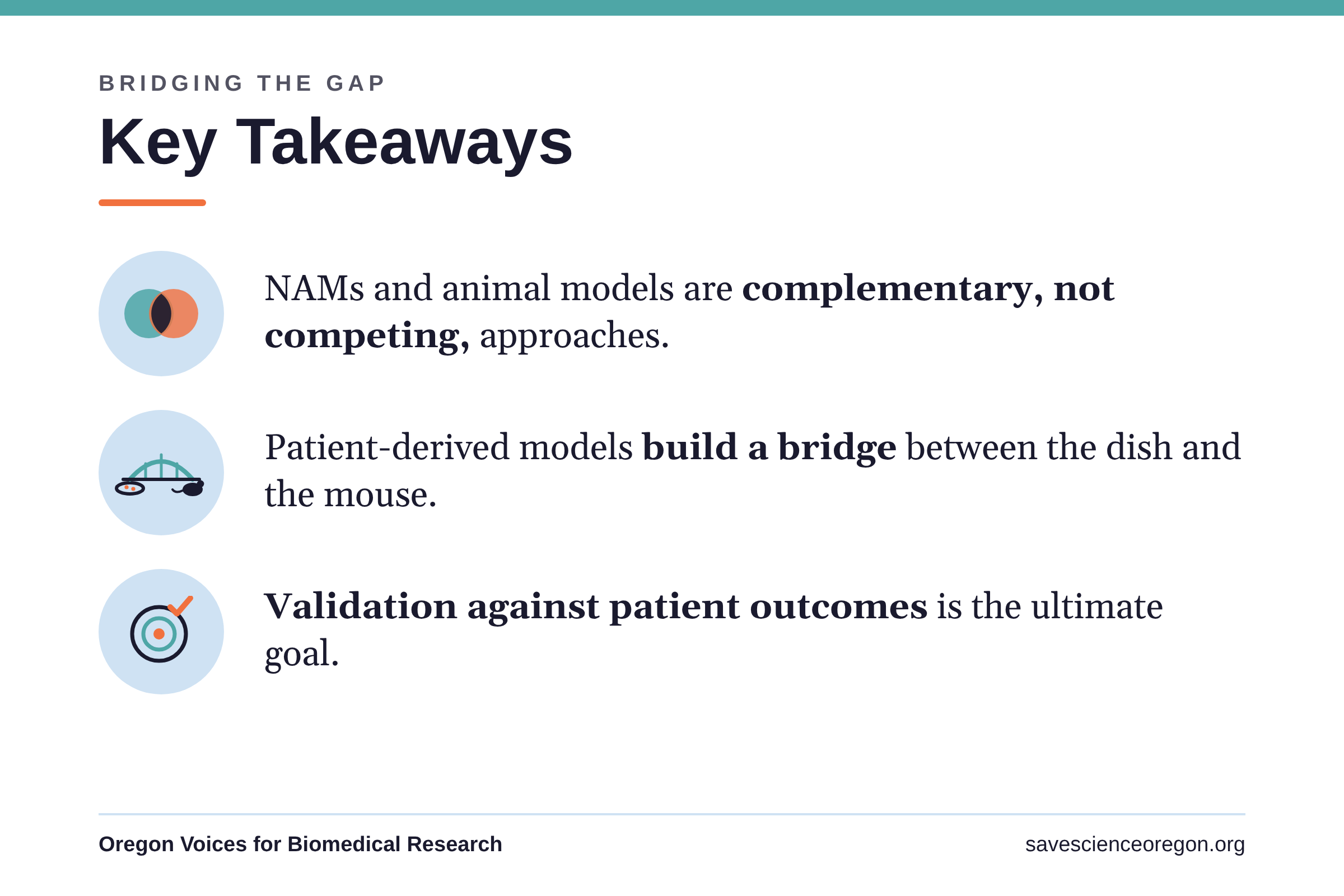
Ultimately, validation is the goal of any research model, and this consortium offers strong examples of how NAMs and animal models can work together toward it. As science and technology advance, and as NAMs become less dependent on animal models, we can expect to reduce the number of animals needed in research. Researchers already work to weigh the validity, integrity, and ethics of whatever model they choose, and we can support those efforts by treating NAMs and animal models as collaborators, not competitors.
References
- Finan, J. M., Di Niro, R., Park, S. Y., et al. (2024). The polymeric fluoropyrimidine CF10 overcomes limitations of 5-FU in pancreatic ductal adenocarcinoma cells through increased replication stress. Cancer Biology & Therapy, 25(1), 2421584. https://doi.org/10.1080/15384047.2024.2421584. PMID: 39513592
- Habowski, A. N., Budagavi, D. P., Scherer, S. D., et al. (2024). Patient-derived models of cancer in the NCI PDMC Consortium: Selection, pitfalls, and practical recommendations. Cancers, 16(3), 565. https://doi.org/10.3390/cancers16030565. PMID: 38339316
- Kuhn, M. R., Wolcott, E. A., & Langer, E. M. (2025). Developments in gastrointestinal organoid cultures to recapitulate tissue environments. Frontiers in Bioengineering and Biotechnology, 13, 1521044. https://doi.org/10.3389/fbioe.2025.1521044. PMID: 40313639
- Langer, E. M., Allen-Petersen, B. L., King, S. M., et al. (2019). Modeling tumor phenotypes in vitro with three-dimensional bioprinting. Cell Reports, 26(3), 608-623.e6. https://doi.org/10.1016/j.celrep.2018.12.090. PMID: 30650355
- Shah, V. M., Rizvi, S., Smith, A., et al. (2023). Micelle-formulated juglone effectively targets pancreatic cancer and remodels the tumor microenvironment. Pharmaceutics, 15(12), 2651. https://doi.org/10.3390/pharmaceutics15122651. PMID: 38139993
- Tessier, J., Grygoryev, D., Chow-Castro, M., et al. (2025). Protocol to purify and culture human pancreatic cancer cells from patient-derived xenografts. STAR Protocols, 6(1), 103672. https://doi.org/10.1016/j.xpro.2025.103672. PMID: 40048424